Figure 1.

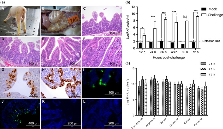
Pathogenicity of PDCoV strain NH (P10). (a) (A) At 28 hr post‐infection, piglets exhibited diarrhoea and yellow faeces were observed behind the legs. (B) Dilated and thin intestinal walls were observed in the piglets terminated at 48 hpi. (C) Pigs were euthanized for pathological examinations at 72 hpi. (D) Severe villous atrophy of the jejunum and ileum was observed. (E) Negative pigs displayed no obvious lesions or pathological changes in the (E) jejunum or (F) ileum. (G, H) PDCoV antigen was detected using IHC and IFA. The jejunum and ileum stained (I) IHC and (J) IFA positive. No PDCoV antigens were detected in the (K) jejunum or (L) ileum in the mock group. (b) Viral RNA copies in faecal swabs and intestine tissues were detected by RT‐qPCR. (c) Viral levels in the jejunum and ileum were higher than in other intestinal sections. Data are expressed as the mean ± SD of triplicate samples. *p < .05; ***p < .001
