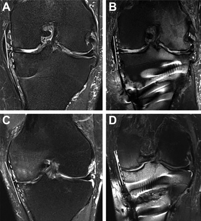
Figure 2.

Preoperative (A and C) and postoperative (B and D) measurements of medial meniscal extrusion: coronal section of meniscus showing the best image of the tibial eminence. A tangent line was drawn from the edge of the medial tibial plateau to the medial femoral condyle (the white dashed line).
