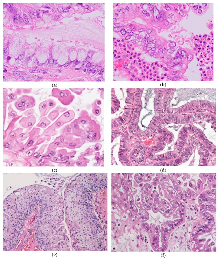
Figure 4.
Seromucinous borderline tumor; microscopic pictures showing a variety of cell types. (a) Sometimes mucinous cells with voluminous cytoplasm can mimic goblet cells. Admixed lightly eosinophilic ciliated cells are almost always identifiable. (b) Background of prominent neutrophilic infiltration. Admixture of mucinous cells, eosinophilic cells, and some clear cells, with mild or moderate nuclear atypia and stratifications. (c) Indifferent cells with abundant eosinophilic cytoplasm. (d) Endometrioid type epithelium. (e) Squamous epithelium. (f) Clear cells. Focal hobnail appearance is also seen (right lower corner).

