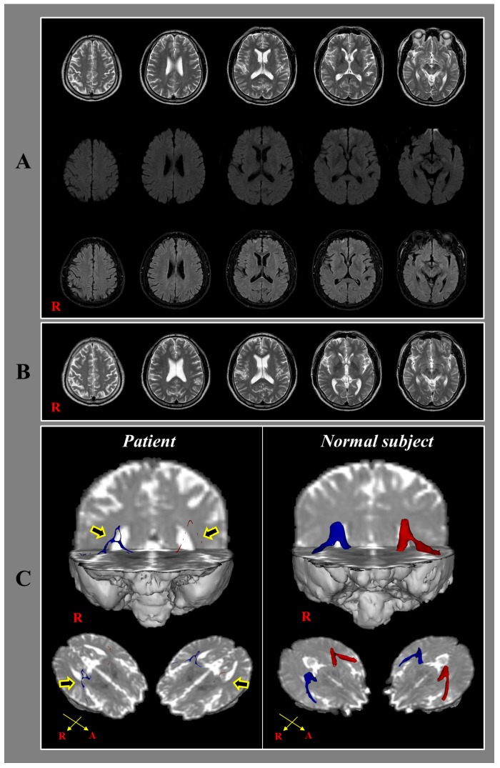
Figure 1.
(A) T2-weighted (upper row), diffusion weighted (middle row) and fluid-attenuated inversion recovery images (lower row) taken 2 weeks after onset reveals no abnormal lesion; (B) T2-weighted images taken 2.5 years after onset shows no abnormal lesion; (C) 2.5-year diffusion tensor tractography for the auditory radiation in the patient shows severe narrowing and tearing (arrows) in both hemispheres compared with a normal subject (47-year old female).

