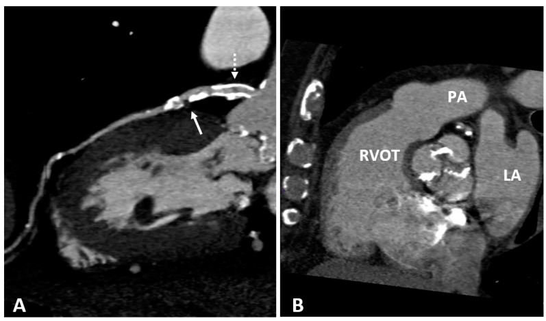
Figure 3.
(A) CTCA showing the left main (LM) and left anterior descending (LAD) coronary arteries of a patient who underwent coronary angioplasty (PTCA) and stent positioning in the LM. A non-calcified low density soft plaque (white arrow) is shown between calcific plaques; the hypodense spot (dashed white arrow) within the stent may indicate initial intrastent restenosis. Calcification of the aortic valve leaflets. (B) CTCA of the same patient reformatted in the plane of the aortic valve shows calcification of the aortic leaflets. RVOT: right ventricle outflow tract; LA: left atrium; PA: pulmonary artery.

