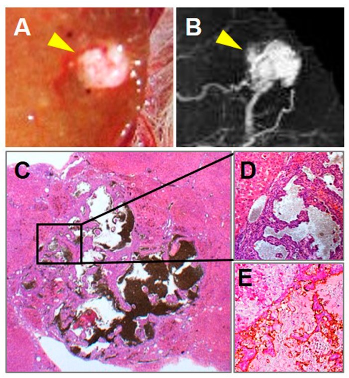
Figure 2.
Pathological specimen, microangiographic and microscopic results of a representative angioma-like HCC. Macrographic (A) and (B) microangiographic views of a tumor-bearing liver lobe revealed a hyper-vascularized lesion (arrowhead) perfused by barium sulfate suspension. Microscopically with H&E staining, the angioma-like HCC was classified as an undifferentiated cancer type (C, original magnification ×12.5, scale bar = 800 µm), filled with enlarged intratumoral vascular lakes, and capsuled or circumscribed by fibrous, on liver cirrhosis background (D. Original magnification ×100, scale bar = 100 µm). CD34-PAS dual staining indicated that tumor blood pools were lined by positively stained endothelia (E. original magnification ×100, scale bar = 100 µm).

