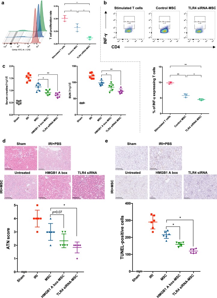
Fig. 6.
HMGB1-TLR4 blocking enhanced immunosuppression and improved MSC-based therapeutics. TLR4 siRNA-treated MSCs and control MSCs were co-cultured with mice spleen-derived CD4+ T cells at a ratio of 1:20 in the presence of IFN-γ, TNF-α (5 ng/ml) and HMGB1 (1 μg/ml) for 72 h. a T cell proliferation was measured by CFSE assay. b The percentage of IFN-γ expressing T cells was assessed to represent the T cell activation. c Differently treated MSCs were administered in mice IRI model. Scr and BUN were detected as indicators of kidney function. d Histological analysis was performed to evaluate kidney structure and quantified by ATN scores. e TUNEL staining was also conducted to evaluate apoptosis. The number of apoptotic cells was counted under light microscope at × 200 magnification. Each data point represented the average value of triplicated wells from each independent experiment (a, b). n = 6 mice per group (c–e). Data were pooled from at least three independent experiments. Data were shown as mean ± SD. *P < 0.05, **P < 0.01

