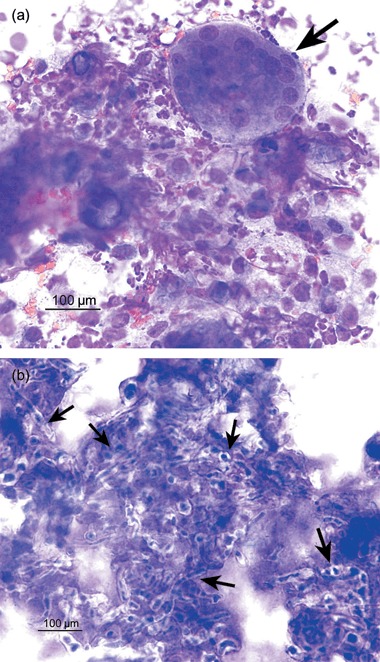
Figure 2.

Stained cytology (Rapi‐Diff II®) of a needle aspirate from the lesion in the Persian cat. Numerous degenerate neutrophils, activated macrophages, multinucleate giant cells (arrow 3a) and fungal hyphae (arrows 3b) are evident. Magnification ×400.
