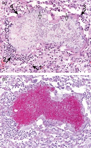
Figure 4.

Histopathology from the Maine Coon's lesion. There is a multifocal, nodular dermatitis with numerous, large, coalescing foci of pyo‐granulomatous inflammation surrounding multiple, irregular, faintly basophilic hyphae‐like elements (open arrows). The inflammatory infiltrate consists mainly of activated macrophages (closed arrow 1), with neutrophils (closed arrow 2), plasma cells (closed arrow 3) and multinucleate giant cells (closed arrow 4). A – haematoxylin and eosin (×200); B – periodic acid Schiff (×200).
