Figure 1.

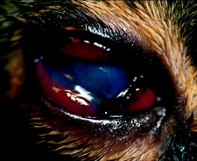
Right eye. Note the mucoid discharge, chemosis and diffuse endothelial corneal edema. The stromal corneal ulceration is behind the nictitating membrane.

Right eye. Note the mucoid discharge, chemosis and diffuse endothelial corneal edema. The stromal corneal ulceration is behind the nictitating membrane.