Figure 2.

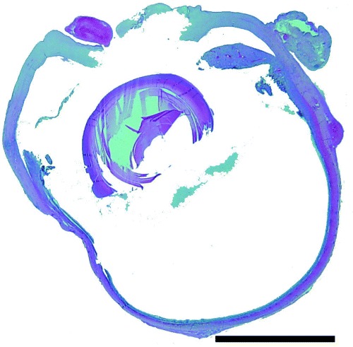
Histopathology of the right globe. Note the central corneal perforation, exudate in anterior chamber and vitreous. Hematoxlyn and eosin. Bar = 2000 µm.

Histopathology of the right globe. Note the central corneal perforation, exudate in anterior chamber and vitreous. Hematoxlyn and eosin. Bar = 2000 µm.