
Figure 5.
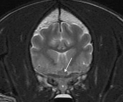
Transverse T2W MR image through the skull of the Golden Retriever at the level of the optic chiasm (arrow) showing diffuse hypointensity of the sphenoid bone relative to normal fatty marrow (*) at first presentation.
Official websites use .gov
A
.gov website belongs to an official
government organization in the United States.
Secure .gov websites use HTTPS
A lock (
) or https:// means you've safely
connected to the .gov website. Share sensitive
information only on official, secure websites.
Transverse T2W MR image through the skull of the Golden Retriever at the level of the optic chiasm (arrow) showing diffuse hypointensity of the sphenoid bone relative to normal fatty marrow (*) at first presentation.