Figure 6.

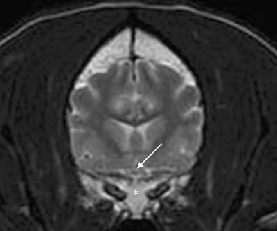
Transverse T2W MR image through the skull of the Golden Retriever at the level of the optic chiasm (arrow) 10 month after first presentation showing the sphenoid bone (*) returned to normal shape and marrow signal.

Transverse T2W MR image through the skull of the Golden Retriever at the level of the optic chiasm (arrow) 10 month after first presentation showing the sphenoid bone (*) returned to normal shape and marrow signal.