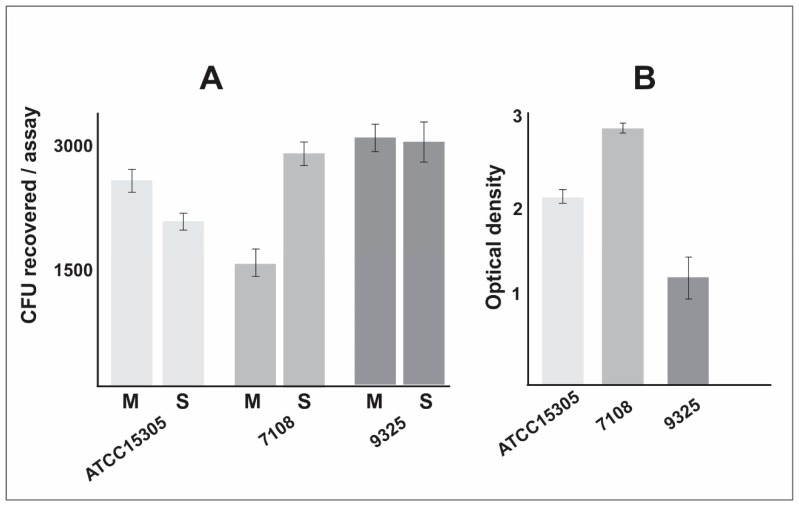
Figure 3.
S. saprophyticus interaction assay with macrophages and evaluation of biofilm formation. (A)The S. saprophyticus cells were incubated with macrophages and, after interaction assay, the supernatant (S) containing non-phagocyted bacterial cells were plated in BHI medium. The macrophages were lysed and colony-forming unit (CFU) recovered and plated in BHI medium (M). The experiments were performed in biological triplicate and standard error of the mean was calculated. (B) The biofilm assay was performed in a polystyrene plate, cells were fixed and stained with crystal violet. Optical density was measured at 570 nm wavelength. The experiments were performed in biological triplicate and standard error of the mean was calculated.

