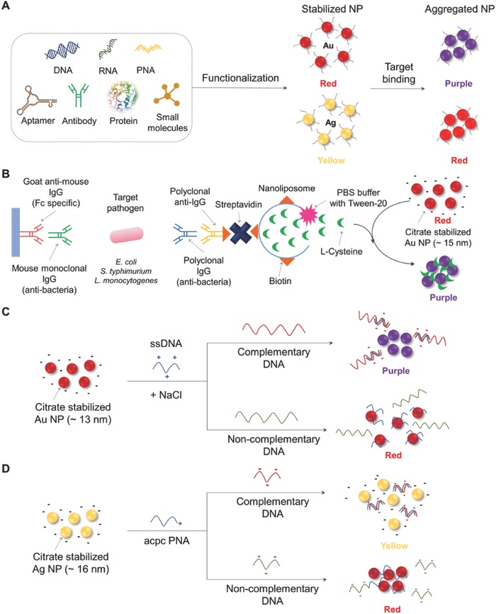
Figure 2.

Schematic illustration of colorimetric detection based on aggregation of NMs. A) Scheme of aggregation of NPs with various biomolecular functionalization. B) Signal‐amplified detection of E. coli, S. typhimurium, and L. monocytogenes using cysteine‐containing nanoliposomes (redrawn from ref. 63). C) Schematic to detect the Cy‐HV3 DNA using aggregation of citrate‐stabilized Au NP in the presence of dsDNA (redrawn from ref. 67). D) Schematic to detect Middle East respiratory syndrome coronavirus DNA using redispersion of Ag NP in the presence of dsDNA (redrawn from ref. 70). PNA: peptide nucleic acid; IgG: immunoglobulin G; PBS: phosphate buffer saline; Cy‐HV3: Cyprinid herpesvirus‐3; ssDNA: single‐stranded DNA; acpc PNA: (2S)‐aminocyclopentane‐(1S)‐carboxylic acid pyrrolidinyl PNA.
