
Figure 2.
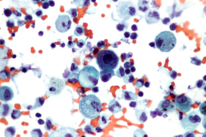
Bronchoalveolar lavage specimen examined after cytocentrifugation (magnification ×40). Besides numerous pulmonary alveolar macrophages and various leukocytes, the specimen revealed a macrophage whose morphologic changes were characteristic of a viral infection with cytomegalovirus. The cell was larger and showed a huge amphophilic intranuclear inclusion with surrounding halo associated to a marked margination of chromatin on the inner surface of the nuclear membrane. The texture of the cytoplasm was enhanced.