
Figure 1.
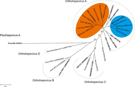
Phylogenetic tree based on nucleotide sequences of complete capsid protein from different hepatitis E virus (HEV) and HEV‐related viruses. Phylogenetic tree was inferred using the maximum likelihood method. A bootstrap analysis of 1000 replicates was included, and the results displayed on the interior branches. The tree is drawn to scale, with branch lengths measured in the number of substitutions per site. Evolutionary analyses were conducted in MEGA6. Sequences in bold represent reference sequence listed by Smith et al., 2014 (Table 1). Accession numbers are shown in brackets. Hepatitis E virus found in bovids and molluscs is not presented here. Genotypes HEV‐3 and HEV‐4 are, respectively, coloured in orange and blue. [Colour figure can be viewed at http://wileyonlinelibrary.com].