Figure 2.

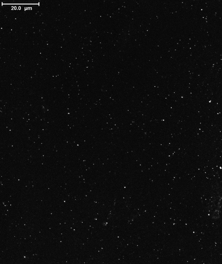
Fluorescence microscopy of VLPs in the bushmeat sample no STE0014. All images were acquired with a Leica SP5 inverted confocal microscope with four lasers, a 100× objective and a numerical aperture of 1.4. The scale bar represents 20 μm.

Fluorescence microscopy of VLPs in the bushmeat sample no STE0014. All images were acquired with a Leica SP5 inverted confocal microscope with four lasers, a 100× objective and a numerical aperture of 1.4. The scale bar represents 20 μm.