Figure 5.
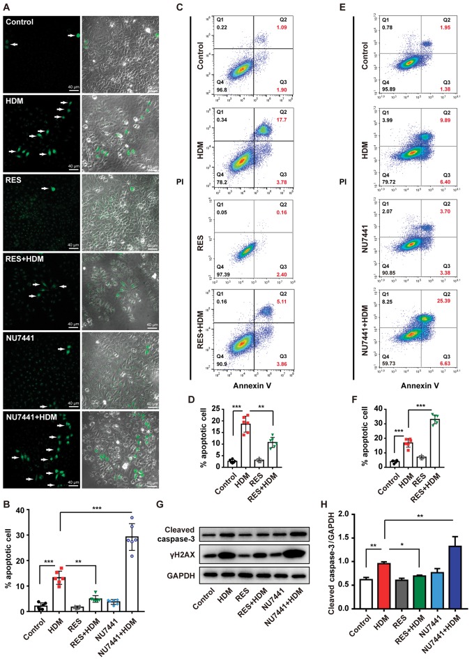
RES protects bronchial epithelial cells from apoptosis induced by HDM treatment. 16HBE cells were incubated with RES or NU7441 for 2 h and then treated with HDM for another 12 h. (A) Representative images of the TUNEL assay which was used to measure apoptosis. (B) Percentage of positive cells in each sample. (C) 16HBE cells were incubated with RES for 2 h and then treated with HDM for another 12 h. Cell apoptosis was detected by flow cytometry with Annexin V-FITC and PI staining and was (D) quantified. (E) 16HBE cells were incubated with NU7441 for 2 h and then treated with HDM for another 12 h. Cell apoptosis was detected by flow cytometry with Annexin V-FITC and PI staining and was (F) quantified. (G) The expression levels of cleaved caspase-3 and γH2AX were determined using western blot analysis. (H) Relative density of cleaved caspase-3 and γH2AX (n=3). Data are presented as mean ± standard deviation. One-way analysis of variance with Tukey-Kramer test or Dunnett's T3 test was used. *P<0.05, **P<0.01 and ***P<0.001. RES, resveratrol; HDM, house dust mites; PI, propidium iodide.

