Figure 4.
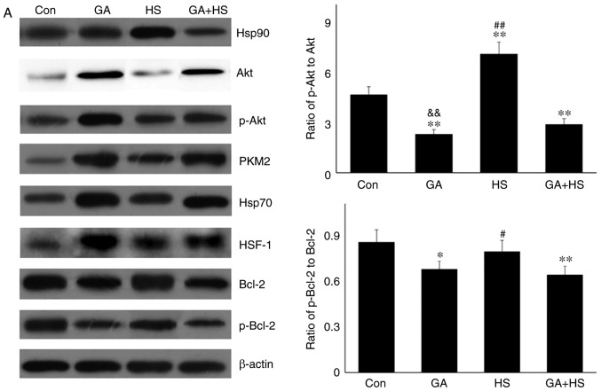
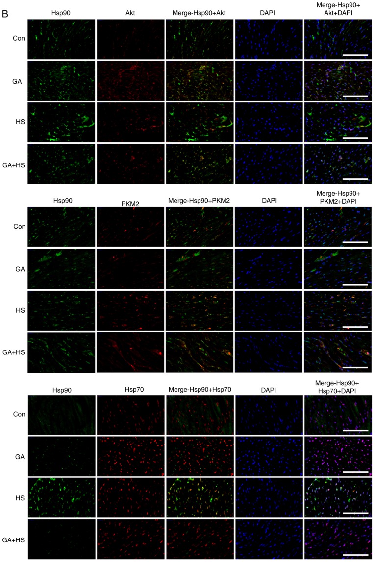
Effect of Hsp90 functional inhibition on the levels of Hsp90, its associated proteins, and the co-localization of Hsp90 with tested client proteins. (A) Representative western blotting results for cellular Hsp90, Akt, p-Akt, PKM2, Hsp70, HSF-1, Bcl-2 and p-Bcl-2 are shown, and the ratios of phos-phorylated/total protein abundance are also presented. (B) Representative immunohistofluorescence staining images showing the co-localization of Hsp90 with Akt, PKM2 and Hsp70 (Hsp90, green fluorescence; Akt/PKM2/Hsp70, red fluorescence; nucleus, blue fluorescence; the merged image of Hsp90 and Akt/PKM2/Hsp70 in the cytoplasm, yellow fluorescence; the merged image of Hsp90 and Akt/PKM2/Hsp70 in the nucleus, white fluorescence) in myocardial tissues. Scale bar=100 µm. The results are expressed as the mean ± SD; n=5. *P<0.05 and **P<0.01 vs. Con, #P<0.05 and ##P<0.01 vs. ASA + HS, and the comparison between ASA and HS is indicated by &&P<0.01.


