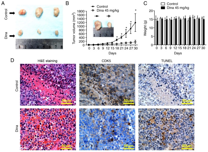
Figure 4.
Anti-myeloma effect of dina in vivo. (A) Representative images of tumor excised from mice on day 30 were shown. (B) Tumor volume was measured every three day. Data shown are mean ± SD. *P<0.05 vs. control. (C) Mouse body weight was measured every three day. Data shown are mean ± SD. (D) Tumor sections from control and dina-treated mice were immunostained with anti-CDK5 antibody (dark brown). Scale bar, 50 µM. Apoptotic cells were identified by a TUNEL assay (TUNEL-positive cells: Dark brown), as well as H&E staining. Photographs are representative of similar observations in three different mice receiving same treatment. SD, standard deviation; CDK, cyclin dependent kinase; H&E, hematoxylin and eosin; TUNEL, terminal deoxynucleotidyl-transferase-mediated dUTP nick end labelling; dina, dinaciclib.

