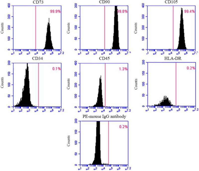
Figure 1.

DPCs phenotype by flow cytometry. The expression of a series of cell surface markers associated with the mesenchymal stem cell (MSC) phenotype was investigated using flow cytometry. Analysis of molecular surface antigen markers in DPCs by flow cytometry indicated that cells were negative for CD34 and CD45, whereas they were positive for CD73, CD90, CD105; PE-conjugated non-specific mouse IgG1 served as negative control.
