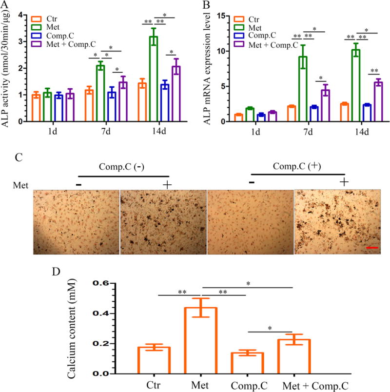
Figure 4.

Effect of metformin-induced ALP activity and mineralized nodule formation in DPCs. (A–B) DPCs were treated with metformin (50 μM) in the absence or presence of Compound C (10 μM, pretreatment for 1 h); cells were retreated every 3 days. ALP activity (A) and ALP mRNA expression (B) were measured at each time point. Data represent mean ± SD of 3 experiments with triplicates. *P < .05. **P < .001. (C) DPCs were cultured in osteogenic induction medium for 14 days, mineralized nodule formation was assessed by von Kossa staining (scale bar = 100 μm). (D) On the 14th day, the calcium content was determined. Data represent mean ± SD of 3 experiments with triplicates. *P < .05. **P < .001.
