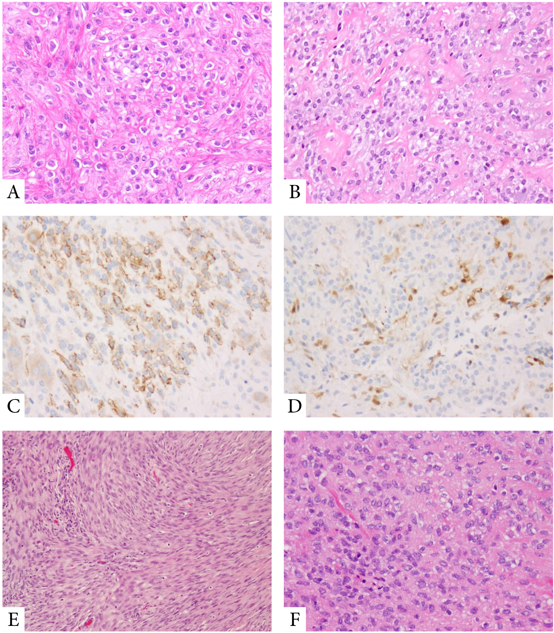
Figure 3. Pathologic features of MET harboring EWSR1-PBX1/3 fusions.

Tumors with EWSR1-PBX1 fusions often show a bland epithelioid to ovoid phenotype, with scant clear cytoplasm, embedded in a delicate fibrous collagenous stroma (A,B). Tumors are frequently positive for EMA (C) and S100 protein (D). MET with EWSR1-PBX3 fusions display a more ovoid to spindle cell appearance, with benign histologic features (E,F).
