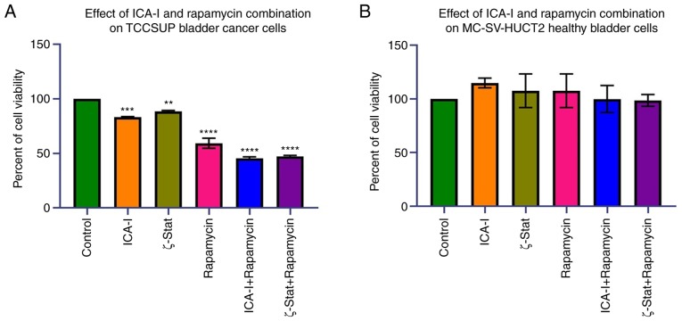
Figure 2.
Effect of atypical PKC inhibitors and rapamycin on normal and malignant bladder cells. (A) TCCSUP and (B) MC-SV-HUCT2 cells (3×10³ cells/well) were plated in 96-well plates and treated with 7.5 μM ICA-I or ζ-Stat, or 100 nM rapamycin, or combination of atypical PKC inhibitor and rapamycin for 72 h. Following incubation with a WST-1 reagent for 3 h, absorbance was determined at 450 nm using a microplate reader. All the experiments were performed for at least three times. Data are presented as the mean ± SEM. **P<0.02, ***P<0.01 and ****P<0.0001 vs. untreated control cells. PKC, protein kinase C.

