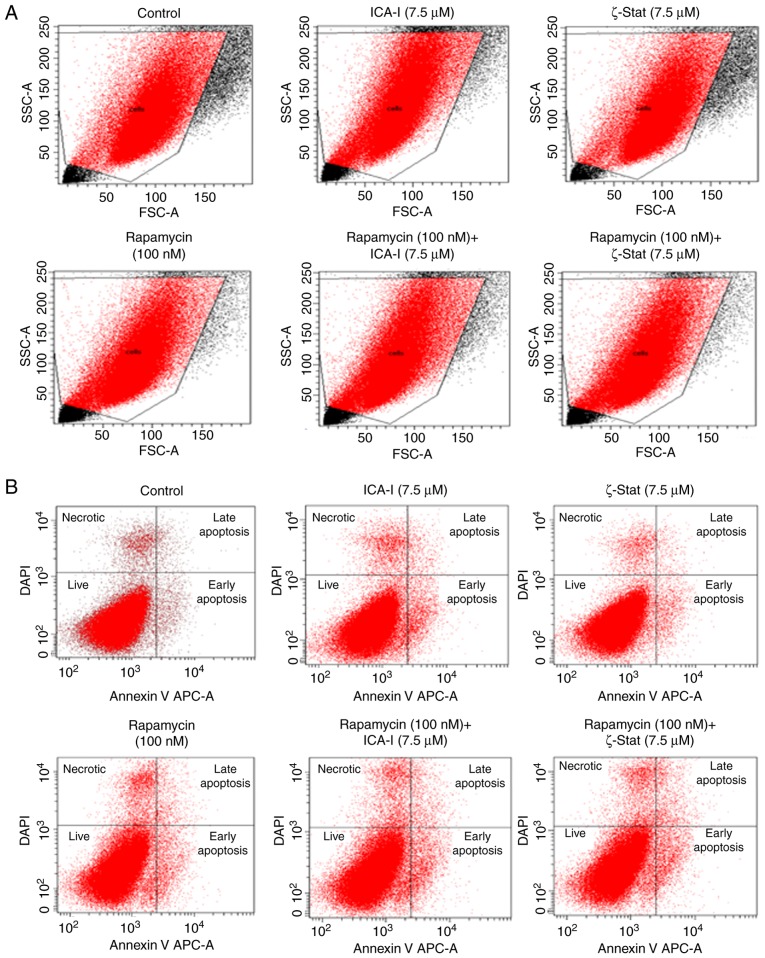
Figure 3.
Effect of atypical PKC inhibitors and rapamycin on malignant bladder cell apoptosis. TCCSUP cells (1×10⁶) were plated in 100-mm plates and treated with 7.5 μM ICA-I or ζ-Stat, or 100 nM rapamycin or combination of atypical PKC inhibitor and rapamycin for 72 h. Following treatment, the cells were harvested and labeled with an APC-bound Annexin V and DAPI to detect different stages of apoptosis using flow cytometry. (A) Targeted cells used for gating during flow cytometric analysis to detect necrotic and apoptotic populations. (B) Percentage of different phases of apoptotic and necrotic population using flow cytometer. The experiment was repeated for N=3 independent times. PKC, protein kinase C; SSC, side scattered light; FSC, forward scattered light.

