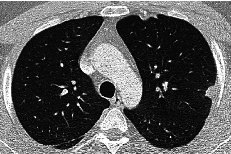
Fig. 1.

Chest HRCT, transverse plane, initial examination. At the aortic arch level, a subpleural nodule in the apical S1+2 segments of the left upper lobe with pleural thickening in the adjacent area. Subpleural lesions are quite typical for necrotizing sarcoid granulomatosis (NSG) without predilection for upper or lower lobe involvement. In the right lung dorsally, a slight thickening of the pleura and of the major interlobar fissure
