
Fig. 3.
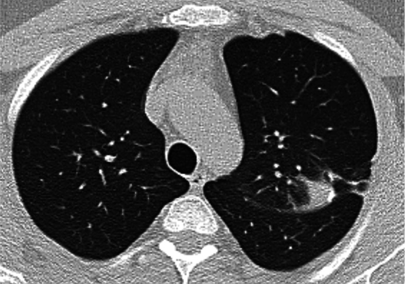
Chest HRCT, transverse plane, two-month follow-up. A tiny ‘consolidation’ of the pulmonary parenchyma with isolated calcifications and fibrous streaks extending towards the thickened pleura is present at the upper left lobe level in S1+2. The original S1+2 nodule adjacent to the pleura has partially regressed – a sign of a certain development of the disease over time