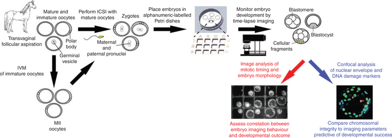
Fig. 1.
Experimental approach to assess equine embryo developmental potential by time-lapse monitoring. Equine oocytes were obtained by transvaginal aspiration from cross-bred mares (n = 9) undergoing ovum pick-up and matured in vitro. Mature MII oocytes were fertilised via intracytoplasmic sperm injection (ICSI) and presumed zygotes (n = 56) were placed in alphanumeric-labelled Petri dishes for embryo tracking and non-invasive time-lapse image analysis of preimplantation development to the blastocyst stage. Early mitotic timing and morphological criteria predictive of blastocyst formation were measured and correlated with embryo developmental outcome. Confocal imaging was used to assess nuclear structure and DNA integrity in arrested cleavage stage embryos versus those that reached the blastocyst stage.

