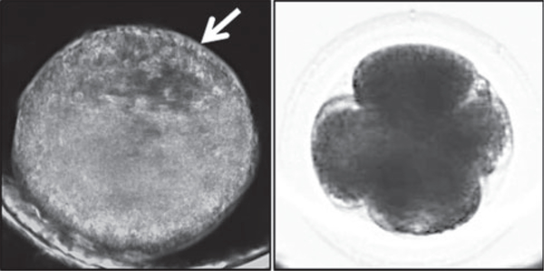
Fig. 2.
Comparison of darkfield and brightfield imaging of equine preimplantation embryogenesis. (a) Image frame of an equine blastocyst monitored by darkfield time-lapse imaging throughout preimplantation development. The white arrow indicates the blastocoel of the blastocyst. (b) A 4-cell equine embryo visualised using the bimodal microscope shows brightfield imaging is more suitable for assessing early mitotic timing.

