Fig. 6.
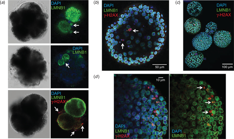
Micronuclei are prevalent and susceptible to DNA damage in equine embryos. (a) Confocal imaging of 4′,6′-diamidino-2-phenylindole (DAPI)-stained (blue) arrested cleavage stage embryos immunolabelled with an antibody against the nuclear envelope marker lamin B1 (LMNB1; green) revealed chromosome-containing micronuclei (top panel) and multinuclei (middle panel). Additional immunostaining with an antibody against the histone variant H2AX phosphorylated at serine 139 (red), a marker of double-stranded DNA breaks, in arrested embryos demonstrated that γ-H2AX-positive foci are associated with micronuclei formation (bottom panel). (b)Similar imaging of DAPI-stained blastocyst stage embryos immunolabelled for LMNB1 and γ-H2AX demonstrated that blastocysts can retain micronuclei. (c) Micronuclei were detected in most blastocysts and exhibited DNA damage, as indicated by the intensity of γ-H2AX immunostaining. (d) The prevalence of DNA breakage in blastocysts was likely due to the lack of, or a defect in, nuclear envelope surrounding micronuclei (white arrows).

