
Figure 7.1.21.
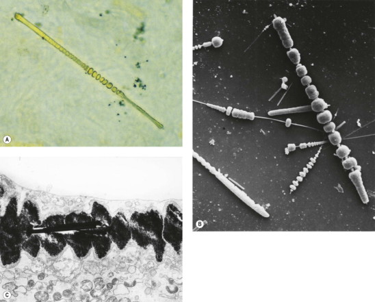
Asbestos bodies seen by light microscopy in (A) an unstained 30-µm-thick paraffin section; (B) by scanning electron microscopy in the digest of an asbestos worker's lung and (C) by transmission electron microscopy in lung tissue. The asbestos fibres have acquired the iron-protein coating that characterises an asbestos body. In most places the coating has become segmented, giving rise to bead-like formations, a change accompanying ageing of the bodies.
((B) Courtesy of Dr B Fox, London, UK and (C) courtesy of Miss A Dewar, Brompton, UK.)