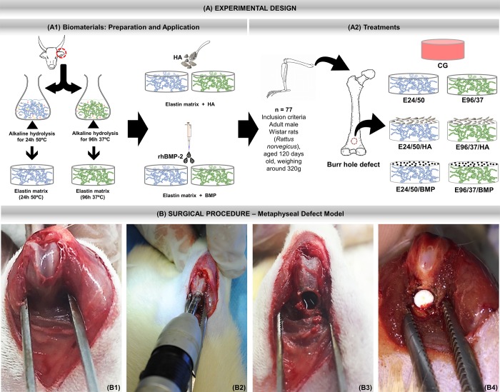
Fig 1.
(A) Experimental design. (A1) Preparation and application of biomaterials. Hydrolysis process of bovine atrial cartilage in alkaline solution at 50°C for 24 hours and 96 hours at 37°C. After obtaining the elastin matrices, either synthetic hydroxyapatite (HA) ceramics or human recombinant morphogenetic protein two are incorporated. (A2) Treatments. Inclusion criteria: 77 male rats, Rattus norvegicus, Wistar, 120 days old and average weight of 320 grams were randomly separated into 7 groups.: CG—Control group (n = 11)–metaphyseal defect filled with blood clot; E24/50 (n = 11)—hydrolyzed elastin membrane filled metaphyseal defects for 24h at 50° C; E24 /50/HA (n = 11)—hydrolyzed elastin membrane filled metaphyseal defects for 24h at 50°C associated with hydroxyapatite; E24/50/BMPP (n = 11) -metaphyseal defects filled with elastin membrane hydrolyzed for 24h at 50°C associated with BMP; E96/37 (n = 11)—hydrolyzed elastin membrane filled metaphyseal defects for 96h at 37°C; E96/37/HA (n = 11)—Elastin membrane filled metaphyseal defects hydrolyzed for 96h at 37°C associated with hydroxyapatite; E96/37/BMP (n = 11)—metaphyseal defects filled with elastin membrane hydrolyzed for 96h at 37°C associated with BMP. (B) Surgical procedure—Metaphyseal Defect Model. B1) Medial parapatellar incision in the right femur followed by arthrotomy, lateral patellar dislocation and visualization of the femoral condyles. (B2) Metaphyseal Defect Model made with 3 mm diameter trephine drill. (B3) Circular osteotomy in the anterior metaphyseal region of the distal femur. (B4) Defect filled with elastin matrix.

