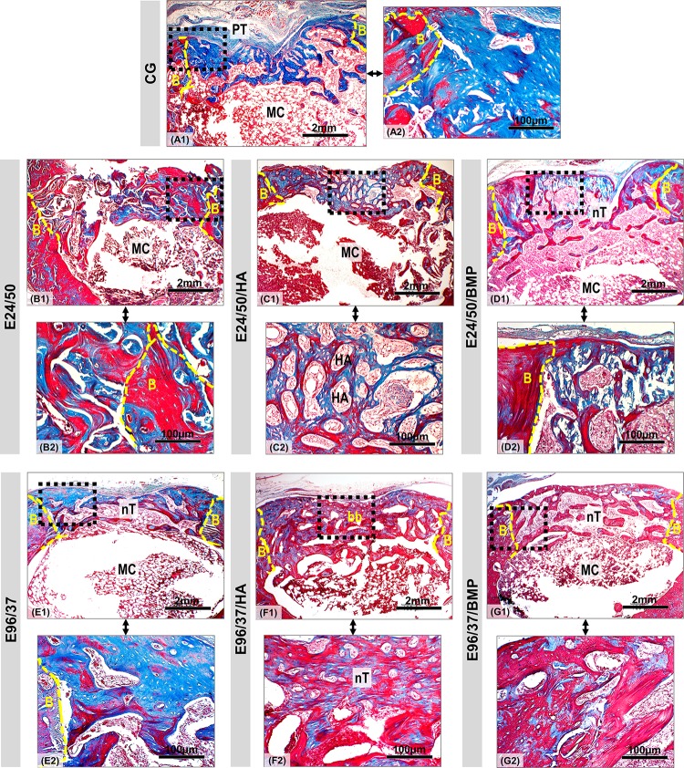
Fig 9. Panoramic view of bone defect created in distal femoral metaphysis in rats.
Defect area in the CG, E24/50, E24/50/HA, E24/50/BMP, E96/37, E96/37/HA and E96/37/BMP (A1-G1) experimental groups at 42 days. All experimental groups presented trabecular bone formation from the remnant cortical border in transition from immature to mature bone, but without complete healing of the surgical wound. In the E96/37/HA treated defect, the bone trabeculae were more mature, thicker and more organized, forming a bone bridge connecting the defect edges. Masson's trichrome staining: new bones with blue dye and mature bones with red dye. Periosteal tissue (PT); defect border (B); medullary canal (MC); neoformed bone trabeculae in the cortical region (nT); bone bridge (bb); Hydroxyapatite particle (HA). Masson's trichrome staining of defect area. Original 4x magnification, Bar: 2mm. Inset 40x magnified images (A2-G2). Bar: 100 μm.

