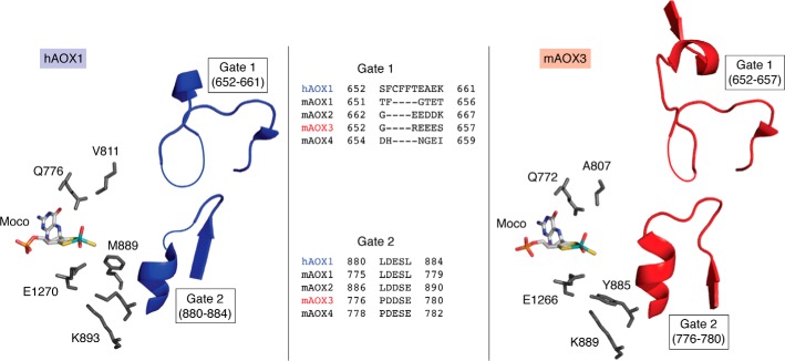
Figure 7.
The active site of hAOX1 and mAOX3. Shown are the residues of hAOX1 (left) and mAOX3 (right) surrounding the Moco and the location of Gate 1 and Gate 2 at the entrance of the substrate funnel. Missing residues in the electron density (and therefore not present in the coordinate files) are indicated by thin lines. An amino acid sequence alignment of Gate 1 and Gate 2 of the human and mouse enzymes is given. The figure was created using PyMOL version 2.1.1.

