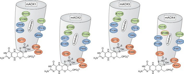
Figure 8.
Active site and substrate binding funnel in mouse AOX enzymes. Representation of the substrate binding sites in mouse AOX1, AOX2, AOX3, and AOX4 in addition to residues in the conserved and nonconserved substrate-binding funnel, which selects the substrate specificity. The representation of mAOX3 is taken from the crystal structure (PDB: 3ZYV) (38), whereas those of mAOX1, mAOX2, and mAOX4 are taken from the modeled structures (54). Residues in red indicate the amino acids whose nature is conserved in all mouse AOX enzymes; residues in blue are hydrophobic residues, partially conserved and involved in substrate orientation; and residues in green are those specific for AOX4, with the AOX4-Met1088 residue highlighted in red. The funnel for mAOX4 is predicted to be smaller compared with the other enzymes.

