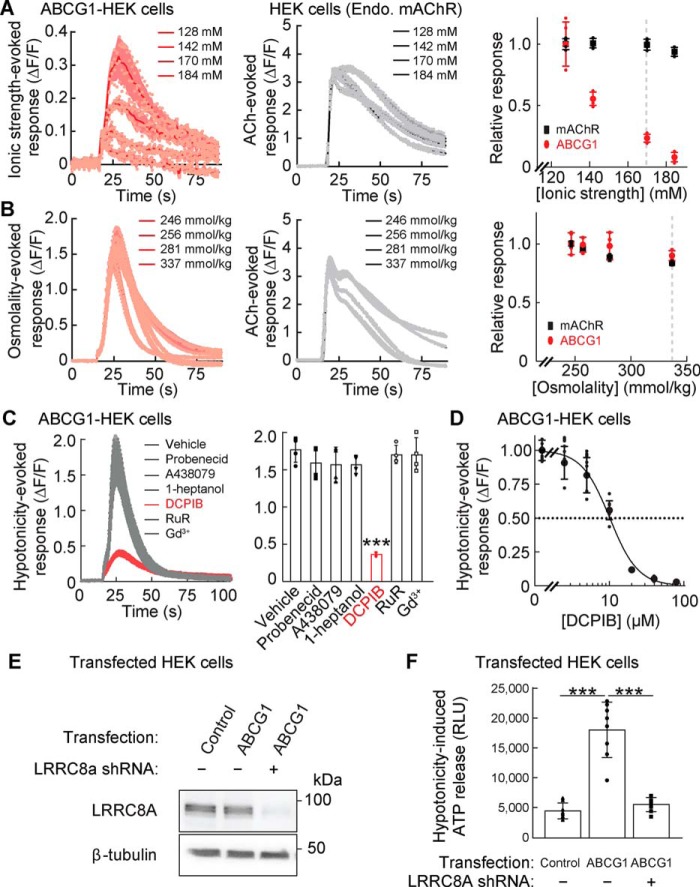
Figure 4.
Ionic strength-evoked VRAC activity mediates hypotonicity-induced ATP release. A–D, stimulus-induced calcium responses were measured from ABCG1- or RFP (control)–transfected HEK cells with the calcium FLIPR assay. A, lower ionic strength induced larger calcium responses from ABCG1-transfected HEK cells but did not alter acetylcholine (ACh)–induced (final concentration, 100 μm) calcium responses of endogenous mAChR in RFP-transfected HEK cells (n = 4). Solutions with various ionic strengths maintain a constant osmolality (340 mmol/kg) with mannitol. B, solutions with various osmolalities were prepared while maintaining a constant ionic strength (final concentration, 113 mm). The osmolality solutions without and with ACh (final concentration, 100 μm) similarly induced calcium responses from ABCG1-transfected cells and HEK cells, respectively. Osmolality had no obvious effects on peak calcium responses (n = 4). C, hypotonic stimulation (final concentration, 250 mmol/kg) induced calcium responses (ΔF/F) from ABCG1-transfected HEK cells, which were decreased significantly by 1 h of preincubation with the VRAC inhibitor DCPIB (final concentration, 20 μm) but not with inhibitors of pannexin (final concentration, 2.5 mm probenecid), P2X7 receptor (final concentration, 5 μm A438079), connexin (final concentration, 1 mm heptanol), CALHM (final concentration, 20 μm ruthenium red (RuR)), and Maxi anion channel (final concentration, 50 μm Gd3+) (n = 4) (one-way ANOVA; F(6,21) = 31.97; p < 0.001). D, hypotonicity (final concentration, 250 mmol/kg)–induced peak calcium responses (ΔF/F) were measured in ABCG1-transfected HEK cells preincubated with various doses of DCPIB (IC50 = 10.2 ± 0.4 μm) (n = 8). E and F, ABCG1 does not alter LRRC8A expression. LRRC8A-deficient or control HEK cells were transfected transiently with RFP (control) or ABCG1. E, whereas the loss of LRRC8A protein was confirmed in LRRC8A-deficient HEK cells, ABCG1 transfection did not alter LRRC8A protein amount. The β-tubulin protein amount was unaltered under all conditions examined. F, ABCG1 transfection increased hypotonicity (final concentration, 250 mmol/kg)–induced ATP release in the control HEK cells, but not in the LRRC8A-deficient HEK cells (n = 8) (one-way ANOVA; F(2,21) = 55.78; p < 0.001). The data are means ± standard deviation (one-way ANOVA followed by Tukey's test (C and F)). ***, p < 0.001. RLU, relative luminescent unit.

