Figure 8.
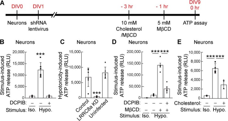
Neurons release ATP upon hypotonic stimulation through cholesterol-dependent VRAC. Primary cerebellar neurons were prepared, and extracellular ATP in the medium was measured 30 s after conditioned Hanks' balanced salt solution (isotonic 340 mmol/kg; Iso.) or hypotonic (final concentration, 250 mmol/kg; Hypo.) stimulation by using the luciferin–luciferase bioluminescence assay. A, experimental overview. B, hypotonic, but not isotonic, stimulation increased extracellular ATP in the neuronal medium, and DCPIB (20 μm) inhibited hypotonicity-induced ATP release (n = 8) (one-way ANOVA; F(2,15) = 74.61, p < 0.001). C, neuronal LRRC8A is required for hypotonicity-induced ATP release. Primary cerebellar neurons were infected with lentivirus carrying LRRC8A shRNA or random shRNA encoded in pFUGW-H1 (control). ATP release upon hypotonic stimulation was abolished in LRRC8A shRNA-infected neurons but was unaltered in uninfected neurons or neurons infected with a control virus (n = 6) (one-way ANOVA; F(2,15) = 24.29, p < 0.001). D, cholesterol depletion by preincubation of neurons with 5 mm MβCD robustly enhanced hypotonicity-induced ATP release, and this enhancement was blocked by the addition of 20 μm DCPIB (n = 8) (one-way ANOVA; F(3,20) = 131.2, p < 0.001). E, cholesterol repletion by preincubation of neurons with a mixture of 5 mm cholesterol with MβCD suppressed hypotonicity-induced ATP release (n = 4) (one-way ANOVA; F(2,9) = 55.95, p < 0.001). The data are means ± standard deviation (one-way ANOVA followed by Tukey's test (B–E)). ***, p < 0.001. RLU, relative luminescent unit.

