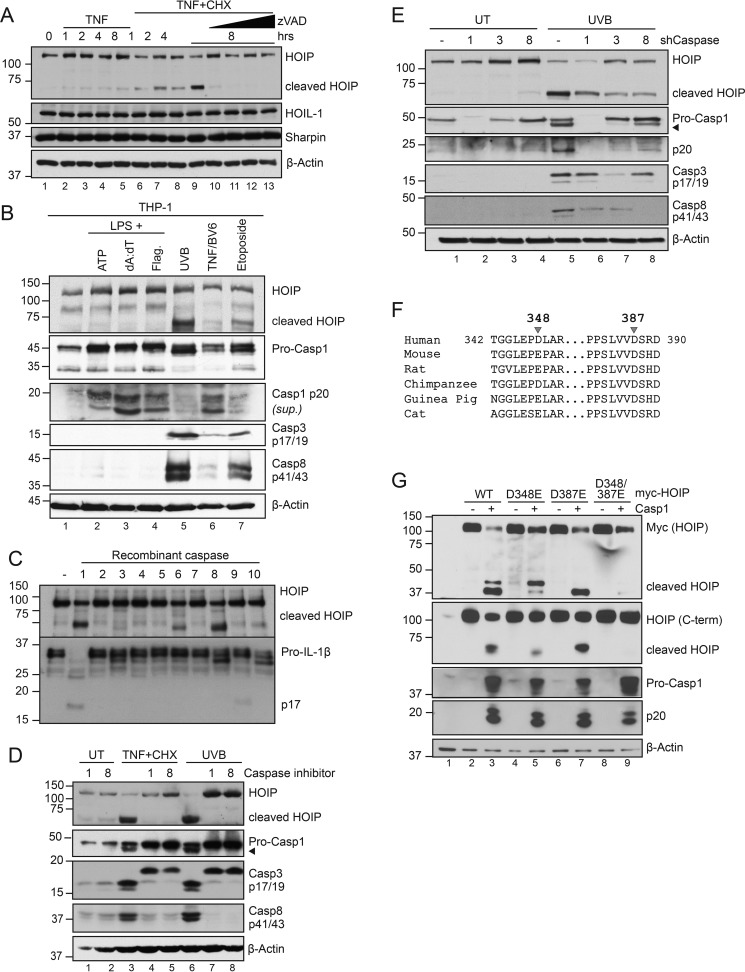
Figure 3.
HOIP is cleaved at Asp-348 and Asp-387 by multiple caspases during apoptosis. A, HaCaT cells were pretreated with CHX (6 μg/ml) with or without Z-VAD (2.5, 5, 10, and 20 μm) for 45 min, followed by TNF (100 ng/ml) for the indicated times. Cells were lysed and subject to immunoblot analysis. Data are representative of two independent experiments. B, PMA-differentiated THP-1 cells were primed with 100 ng/ml LPS for 4 h followed by ATP (5 mm, 1 h), Lipofectamine-transfected poly(dA:dT) (1.5 μg/ml, 6 h), or DOTAP-transfected flagellin (2 μg/ml, 6 h) to engage inflammasomes or with UVB (50 mJ/cm2, 6 h), TNF (100 ng/ml) + BV6 (5 μm) (6 h), or etoposide (200 μm) to engage apoptosis. C, in vitro transcribed and translated HOIP and pro-IL-1β were incubated with a panel of recombinant caspases for 1 h at 37 °C and subjected to immunoblot analysis following reaction termination with Laemmli buffer. 2 units of recombinant caspase was used per reaction (Biovision K233). Data are representative of two independent experiments. D, HaCaT cells were pretreated with YVAD (10 μm; Casp1 inhibitor), IETD (10 μm; Casp8 inhibitor), or CHX (6 μg/ml) for 45 min before treatment with either TNF (100 ng/ml) or UVB (50 mJ/cm2) for 6 h. Cells were lysed and subjected to immunoblot analysis. Data are representative of three independent experiments. E, shCaspase-1, -3, or -8 HaCaT cells were stimulated with 50 mJ/cm2 UVB, lysed 6 h post-irradiation, and subjected to immunoblot analysis. Data are representative of two independent experiments. F, sequence alignment of HOIP (RNF31) from various species. G, HEK293T cells were transfected with equal amounts of the indicated plasmids. 24 h post-transfection, cells were lysed and subjected to immunoblot analysis. Data are representative of two independent experiments.

