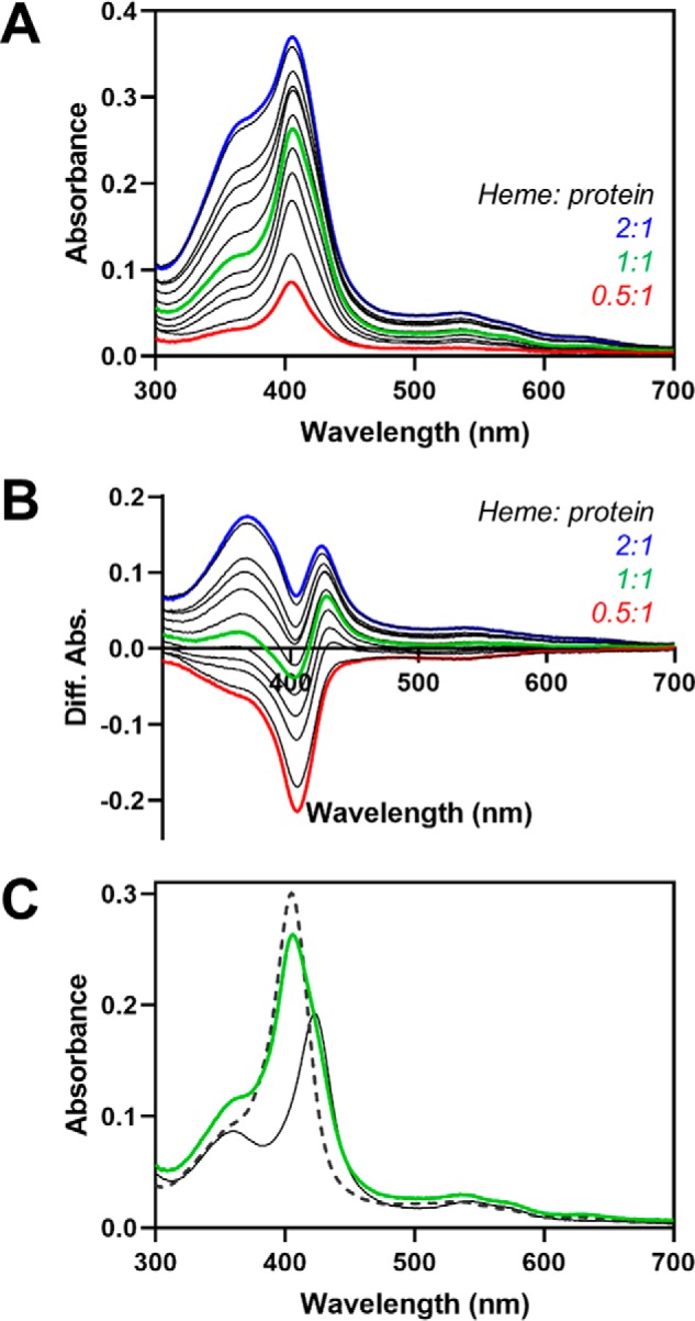
Figure 7.

Fe3+-heme equilibration between HRM1 and the core in C282A HO2solR. Increasing concentrations of heme were incubated with C282A HO2solR in an anaerobic chamber. A, after reaching equilibrium, UV-visible spectra were recorded. B, difference spectra were generated by subtracting the absorbance of heme-bound C282A HO2tail (solid black spectrum in C). C, absorbance spectrum from the same assay in A in which the added heme concentration is equal to the protein concentration of C282A HO2solR (green). The spectra of heme-bound C282A HO2tail (solid black) and heme-bound HO2core (dashed black) are shown for reference and were used to calculate that ∼70% of the added heme is bound to the core.
