Figure 9.
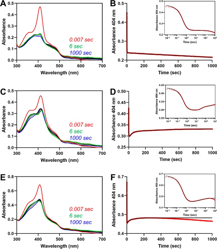
Fe3+-heme transfer from HRM1 to the core after single turnover of heme in the core. A, heme-bound HO2solO (5 μm final concentration) and an equimolar concentration of CPR were rapidly mixed with a limiting amount of NADPH to initiate degradation of heme at the core. All measurements were carried out at 20 °C using the 1-cm path length configuration in photodiode array mode in triplicate. The stopped-flow trace at 404 nm (B, red) was fit to a triple-exponential equation (B, black) using the Pro-data Viewer software provided by Applied Photophysics. The inset is a semi-log plot of the data in B with the data shown in red and the fit in black. C and D, same as A and B except C282A Fe3+core/HRM-HO2solR was used in the assay in place of HO2solO, and data were fit to a quadruple-exponential equation. D–F, same as A and B except that an equimolar concentration of free heme was added to the heme-bound HO2solO and CPR solution before mixing with NADPH. Data were fit to a quadruple-exponential equation.

