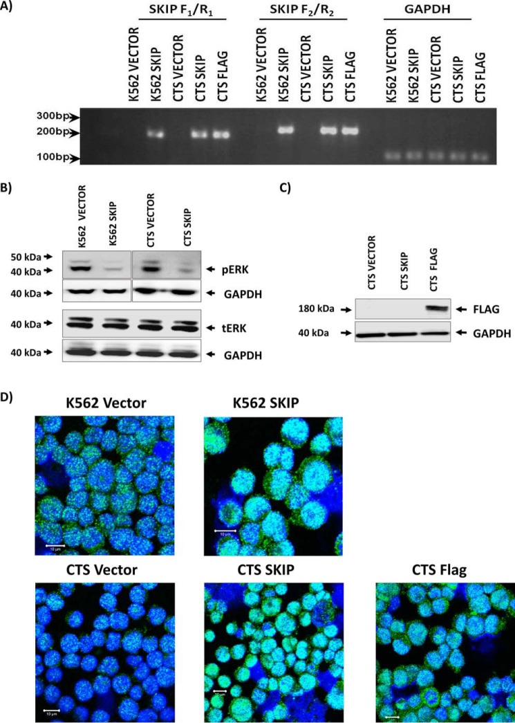
Figure 2.
Successful transfection and re-expression of SKIP protein in K562 and CTS cell lines. A, SKIP gene expression in SKIP-transfected cells was confirmed by RT-PCR using two different primers (F1/R1 and F2/R2), GAPDH expression was used as control for loading. B, Western blots show low pERK phenotype associated with SKIP re-expression in K562 and CTS cell lines and equal tERK expression in the two cell lines. C, Western blotting confirming the expression of FLAG-tagged SKIP protein in FLAG-tagged transfected CTS cell line. D, immunofluorescent detection of SKIP using anti-SKIP antibody and confocal microscopy in SKIP-transfected versus vector alone-transfected K562 and CTS cell lines. Fluorescence was detected using a Zeiss LSM 510 META, confocal microscope system. All immunofluorescent images were taken at a magnification of ×40, scale bar = 10 μm.

