Figure 3.
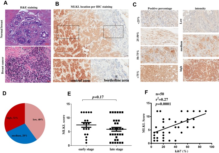
Necroptosis activation is an independent predictive factor for breast cancer malignancy. (A) H&E staining of a breast cancer tissue and a normal breast tissue. (B) immunohistochemistry analysis of MLKL in breast cancer tissues. The subcellular location of MLKL was visualized and its staining intensity in central zone and borderline areas were outlined and magnified, respectively. (C) Representative images of MLKL staining scores. (D) After scoring of MLKL staining, MLKL was classified as low expression, medium expression and high expression. (E, F) The correlation of MLKL expression scores with breast cancer stages and Ki67.
Abbreviations: MLKL, mixed lineage kinase domain-like protein; H&E, hematoxylin and eosin staining; IHC, immunohistochemistry.

