. 2020 Apr 7;9:e55159. doi: 10.7554/eLife.55159

Figure 1. Cap-G expression in Drosophila neurons.

(A) Schematic representation of the Drosophila condensin I complex. (B) w1118 embryo (stage 15, anterior top). Cap-G mRNA is ubiquitously present in neuronal cytoplasm, neurons marked by Elav. (C) Cap-GEGFP embryo (stage 14, anterior top). Cap-GEGFP co-localises with Lola-N in neuronal nuclei of the VNC. (D) Optic lobe of 3rd instar larvae Cap-GEGFP. Cap-G is strongly present in NSCs marked by Dpn. Cap-G is present in neurons (Elav positive) in proximity of NSCs (yellow arrowheads). Scale bars 10 µm.

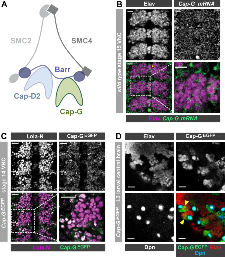
Figure 1.

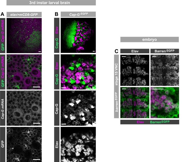
Figure 1—figure supplement 1. Cap-GEGFP expression in neurons.

Figure 1—figure supplement 1.

(A) Optic lobe of 3rd instar larvae. Cap-G mRNA is ubiquitously expressed in the lobes. mCD8-GFP marks neuronal membrane. Zoom in of central brain section shows expression of Cap-G mRNA in neurons marked by elav-driven GFP expression. (B) Optic lobe of 3rd instar Cap-GEGFP larvae. Cap-G is strongly present in the Optic Proliferation centre and the Central brain (CB). Zoom in on CB shows Cap-G is present in neuronal nuclei as it overlaps with marker Elav. (C) BarrenEGFP embryo (stage 15). BarrenEGFP is present in all cells of the VNC, including Elav positive neurons. All images show ventral view of embryonic VNC (anterior -top). Scale bars = 10 µm.