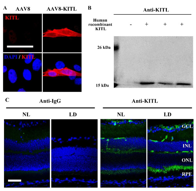
Author response image 2. Analysis of the anti-KITL antibody in photoreceptor cells.
(A) Immunostaining images of anti-KITL in 661W photoreceptor cells infected with AAV8 (left panel) or AAV8-KITL (right panel) virus. (B) Western blots show the specific recognition of the anti-KITL antibody to Human recombinant KITL protein (18 kDa). (C) Immunostaining images of Goat anti-IgG (left panels) and Goat anti-KITL (right panels) in the retinas from 2-month-old albino mice kept under normal light or LD (15, 000 Lux) condition for 1 day. GCL, ganglion cell layer; INL, inner nuclear layer; ONL, outer nuclear layer. Bar: 50 μm.

