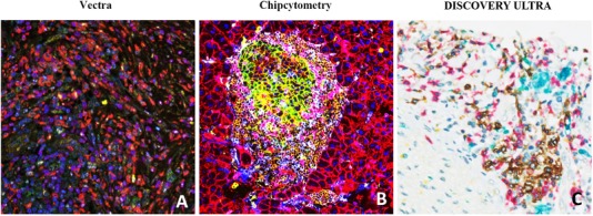
FIGURE 2.

Representative mIHC/IF images captured through the Vectra, Chipcytometry, or DISCOVERY ULTRA imaging system. (A) mIHC/IF of pancreatic adenocarcinoma FFPE sections labelled with DAPI (blue), CD73 (green), CD8 (yellow), CD68 (red), FoxP3 (cyan), CD3 (magenta) and CK (orange) were scanned using the Vectra imaging system. (B) Mouse pancreas FFPE sections labelled with CD45 (brown), CD274 (green), CD3e (purple), CD4 (cyan), CD8a (pink), CD11b (yellow), CD31 (dark brown), CD326/EpCAM (red), B220 (orange), F4/80 (blue), NK1.1 (purple), Pan‐CK (maroon), Hoechst 33342 (dark blue) were scanned using the Chipcytometry imaging system. (C) Cholangiocarcinoma FFPE sections labelled with CD20 (blue), CD8 (red), CD68 (turquoise), CD3 (yellow) were scanned using the DISCOVERY ULTRA imaging system. Abbreviations: mIHC/IF, multiplex immunohistochemistry/immunofluorescence; FFPE, Formalin‐Fixed Paraffin‐Embedded; DAPI, 4′,6‐diamidino‐2‐phenylindole
