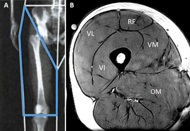
Fig. 1.

a Example of dual-energy X-ray absorptiometry (DXA) image showing regions of interest of the thigh. b Magnetic resonance imaging (MRI) image of the thigh muscles. VI vastus intermedius, VL vastus lateralis, VM vastus medialis, RF rectus femoris, OM other muscles
