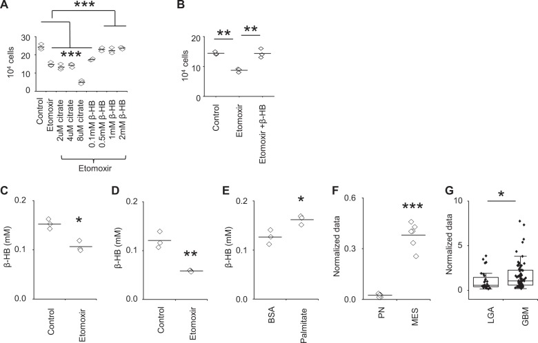
Fig. 3. MES cells produce β-HB in nutrient-rich conditions.
a MES83 cells were treated with +/− etomoxir (40 µM) or in combination with indicated concentration of citrate or β-HB (n = 3). Viable cells were counted using trypan blue after 48 h. b 1027 A cells were treated with +/− etomoxir (40 µM) or in combination with β-HB (0.5 mM) (n = 3). Viable cells counted using trypan blue after 48 h. Cellular β-HB was measured in c MES83 and d 1027 A cells treated with +/− etomoxir (40 µM; 8 h; n = 3). e Cellular β-HB was measured in MES83 cells treated with BSA and BSA:palmitate (500 µM; 4 h; n = 3). Concentration of β-HB in f two patient-derived mesenchymal (MES) and 2 proneural (PN) GBM stem cell lines (n = 3) and g in patient low-grade astrocytoma (LGA; n = 28) and glioblastoma (GBM; n = 80, data truncated at 10). The boxes represent interquartile range, median and whisker denotes upper and lower limit. Line between the data points represents mean. *p < 0.05; **p < 0.005; ***p < 0.0005.

