Fig. 1.
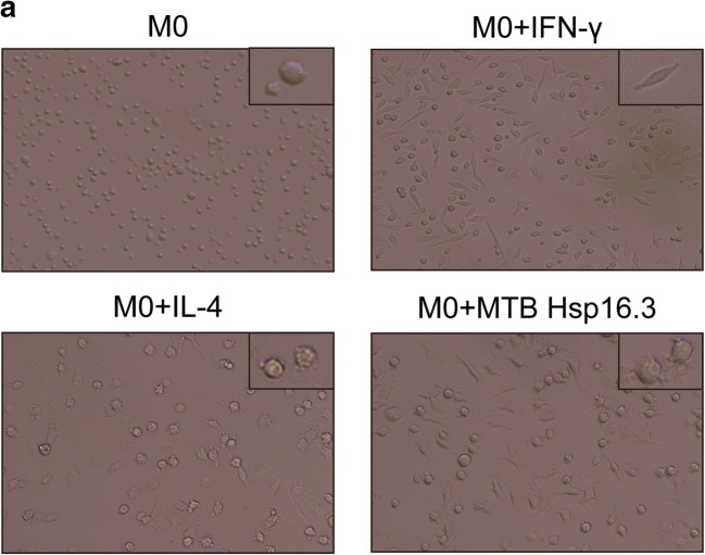
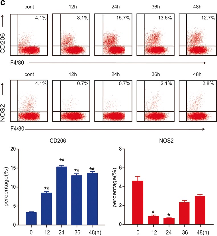
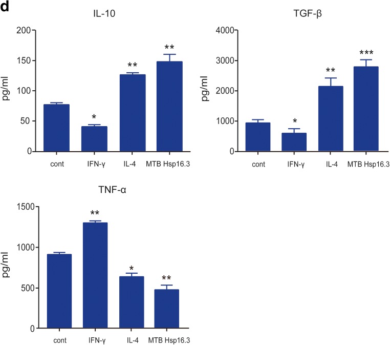
MTB Hsp16.3 induces mouse bone marrow-derived macrophage (BMDM) M2 polarization. Bone marrow cells were isolated from the tibias and femurs of BALB/c mice (6–8 weeks old) and incubated with 20 ng/ml GM-CSF for 7 days. Then, BMDMs were treated with 100 ng/ml MTB Hsp16.3, 100 ng/ml IFN-γ, or 100 ng/ml IL-4 for 0–72 h. a The morphology of BMDMs incubated with IFN-γ, IL-4, or MTB Hsp16.3 for 12 h. The images were captured under an inverted microscope (× 200); the picture in the upper right corner is × 400. Total RNA was extracted from the cells using TRIzol according to the manufacturer’s instructions. b The mRNA expression levels of iNOS, IL-6, TNF-α, Arg-1, IL-10, and TGF-β in macrophages by RT-PCR. c The percentage of F4/80 and NOS2 or CD206 double-positive macrophages was measured by FCM. d The production of TNF-α, IL-10, and TGF-β was measured by ELISA. Data are expressed as the mean ± SEM (n = 3). *p < 0.05, **p < 0.01, ***p < 0.001 compared with the control group (0 h group).


