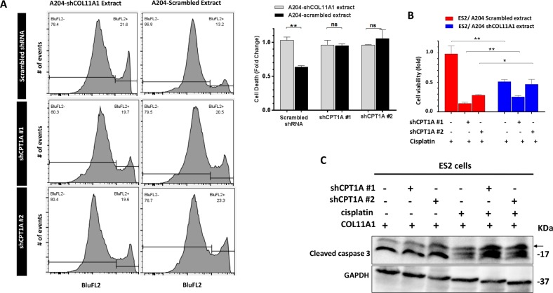
Fig. 5. Inhibition of FAO attenuates COL11A1-induced cisplatin resistance in ovarian cancer cells.
a Flow cytometry analysis of propidium iodide (PI) staining in ES2 cells expressing either scrambled or shCPT1A cultured in COL11A1-positive or negative extract for 48 h and treated for cisplatin (16 µM) for 72 h. Representative flow peak charts are shown on left and quantification is shown on the right. N = 3; y-axis, cell death (fold change); error bar, SD; ns not significant; **p < 0.01. b Relative cell viability of ES2 cells expressing scrambled or shCPT1A vectors cultured in either COL11A1-positive or COL11A1-negative extract and treated with cisplatin (16 µM) for 72 h. N = 3; y-axis, cell viability (fold change); Error bar, SD; *p < 0.05; **p < 0.01. c Western blot of cleaved caspase 3 in ES2 cells expressing scrambled or shCPT1A treated with cisplatin (16 µM) cultured in COL11A1-coated plates for 48 h. GAPDH was used as a loading control.

